MATERIAL DEFECT TROUBLESHOOTING
with Focal Plane Array FTIR (case 05)
The Focal Plane Array (FPA) detector option is currently the most advanced upgrade in our FTIR microscopy lab, which is an excellent add-on for material research and troubleshooting of material defects on micron scale.
QUESTION:
What causes the material defect?
The client requested a failure analysis of an industrial sample that came with an aged 1 μm thick UV-curable coating on a metal substrate. The material defect manifested itself in the pattern areas. These delaminated too easily.
APPROACH:
Focal Plane Array FTIR
The Focal Plane Array (FPA) FTIR method was applied together with SEM/EDX and light microscopy to analyze the material defect.
RESULT:
Unwanted crystallization detected
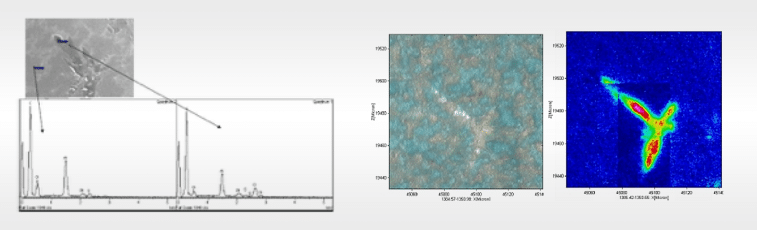
The initial SEM investigation with EDX analysis revealed the presence of local crystallization impeding the curing reaction (figure a). EDX analysis showed accumulation of a Cl-containing species, narrowing down the search to 2 suspicious components.
In addition, other components (with no other elements than CHON) might also have co-crystallized with one of the suspected species as well. With the help of the FPA detector being used in ATR mode we mapped 65535 spectra on a ~ 100×100 μm area within less than 15 minutes.
This ‘spectral cube’ dataset could be analyzed according to the characteristic bands of either of the two components under suspect, creating chemical images for both of them. The analysis clearly revealed the presence of the Cl-containing initiator and the absence of the sensitizer, the other suspicious component.
Figure b (left): light microscope image under polarized conditions.
Figure c (right): stitched FPA FTIR contour plot derived from the 1000 cm-1 band of the Cl-containing initiator.
The IR image (figure c) clearly revealed the defect morphology. That could barely be resolved in the visual image, even under polarized light conditions (figure b). The FPA FTIR mapping also outperformed Raman measurements that confirmed the IR results, but (partial) mapping of the 216 spectra over a 20 x 30 μm area took almost 3 hours.
Want to get to the root of a material defect as well? We can tell you if the Focal Plane Array (FPA) FTIR method is the way to go.
Related Cases


Let's Talk
Fill out this form, and we'll revert to you as soon as possible.
Please provide a detailed description of your question or request.